
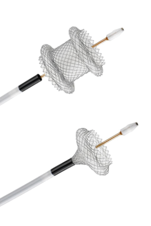
Endoscopic ultrasound (EUS) was initially developed as a diagnostic tool in the 1980s. In 2012, the field of therapeutic EUS expanded significantly due to the introduction of lumen apposing metal stent (LAMS) technology. LAMS are unique dumbbell-shaped metal stents that are deployed under EUS guidance (see image). They enable gastroenterologists with training in interventional endoscopy to connect two lumens and create new routes for drainage, bypasses, and more. Additionally, newer electrocautery-enhanced (or “hot”) devices have transformed LAMS deployment into a single step process, simplifying these new procedures and increasing the margin of safety.
LAMS have caused a paradigm shift in the management of necrotizing pancreatitis. Walled-off pancreatic necrosis (WOPN) can form around four weeks after an episode of necrotizing pancreatitis, and superinfection of these necrotic cavities can be life-threatening. In the past, infected WOPN required surgical necrosectomy and/or percutaneous drains with interventional radiology, both procedures carrying high risks of cutaneous fistula formation. Internal drainage into the stomach or duodenum was possible with endoscopy. However, endoscopic drainage was associated with significant risks, due in part to the lack of ultrasound guidance to assess for vasculature, and the need for multiple steps to achieve drainage of collections. Additionally, these procedures were limited by small caliber plastic stents, which often did not lead to adequate drainage and had issues with getting clogged.
Now, endoscopists can use EUS to identify a safe window to access the cavity, and the 15mm to 20mm lumen of the LAMS can drain necrotic material rapidly. The LAMS is even wide enough for the scope to pass through, so endoscopic necrosectomy can be performed at a later date if needed. Some patients can even have this done as an outpatient! This technique can also be combined with percutaneous drainage to speed up resolution of the cavity. This paradigm change in the management of necrotic collections has obviated the need for surgery in many situations.
Many other applications of LAMS have emerged in the last few years. In carefully selected patients with cholecystitis who are not candidates for surgery, a LAMS can be placed from the duodenum or stomach into the gallbladder, creating durable drainage. Gastric bypass patients with Roux-en-Y anatomy with biliary or pancreatic pathology can have their excluded stomach reconnected with a LAMS to enable ERCP. This technique is also known as EDGE (EUS-Directed transGastric ERCP) and has decreased the need for double balloon assisted ERCP (which has a low success rate) or surgically assisted ERCP (which requires a surgical gastrostomy and can often require prolonged recovery). Endoscopic gastrojejunostomies can be done in select patients with gastric outlet obstructions using LAMS as well. The applications are numerous, and in the pipeline are newer, smaller LAMS that may enable safer biliary drainage for patients who cannot achieve biliary drainage with traditional ERCP.
Not all patients are candidates for these new techniques, however. Current LAMS are 1cm long, so the target lumen needs to be at least that close for the stent to safely deploy. This means that ascites or peritoneal metastases may prevent some patients from benefitting from LAMS. Alternative methods should always be discussed, and multidisciplinary conversations between primary care, gastroenterology, surgery, interventional radiology and oncology are needed. Still, the next time you see a patient with any of the above, consider reaching out to The Oregon Clinic Gastroenterology to find out if your patient could benefit from therapeutic EUS techniques.